Information Possibly Outdated
The information presented on this page was originally released on January 26, 2016. It may not be outdated, but please search our site for more current information. If you plan to quote or reference this information in a publication, please check with the Extension specialist or author before proceeding.
New surgical procedure shows promise for pets
STARKVILLE, Miss. -- A new technique developed by a Mississippi State University veterinarian may improve the long-term management of obstructive hydrocephalus, or water on the brain, in small animals by reducing the rate of surgical complications.
“A similar technique is now used in human infants because, with traditional shunt placement, they can experience a complication rate of about 35 percent within the first two years,” said Dr. Andy Shores, chief of neurosurgery at the MSU College of Veterinary Medicine. “In small animals, statistics show a complication rate of 25 to 50 percent for traditional shunts, and maybe even higher in very young dogs and cats that have much smaller heads. So we looked for a way to improve the procedure of removing excess fluid off the brain in puppies and kittens.”
Obstructive hydrocephalus happens when spinal fluid accumulates in and around the brain in hollow structures called ventricles instead of being reabsorbed by the body. In both humans and animals, neurosurgeons surgically place shunts, or small, tubular structures, through the skull and into the brain to redirect the fluid to another area of the body--usually the abdomen--to be absorbed.
The disorder is most often present at birth and can be caused by several different factors, including heredity, prenatal infection and traumatic birth. Clinical signs include a large, dome-shaped head caused from the buildup of fluid, gait abnormalities, blindness, sleepiness and inability to housetrain, among others.
Small breeds that are short in stature with wide heads, such as Boston terriers, pugs, Maltese, Chihuahuas and toy poodles, are more frequently diagnosed with obstructive hydrocephalus.
Shunts are enclosed in the body but can collapse or become displaced, infected or clogged, Shores said. Bleeding around the shunt is another potential problem.
“Any of these complications must be corrected with another surgery,” Shores said.
This additional surgery is costly for pet owners. Young patients that are still growing could need surgery with the traditional shunts as often as every one to three years, said Dr. Michaela Beasley, a neurosurgeon and assistant clinical professor at the college.
“The new, modified technique allows the same removal of fluid without the high rate of complications caused by placing a traditional shunt in the body,” Beasley said. “With this procedure, we create an alternative drainage and absorption site for the fluid by making an extra opening in the ventricle for the drainage.”
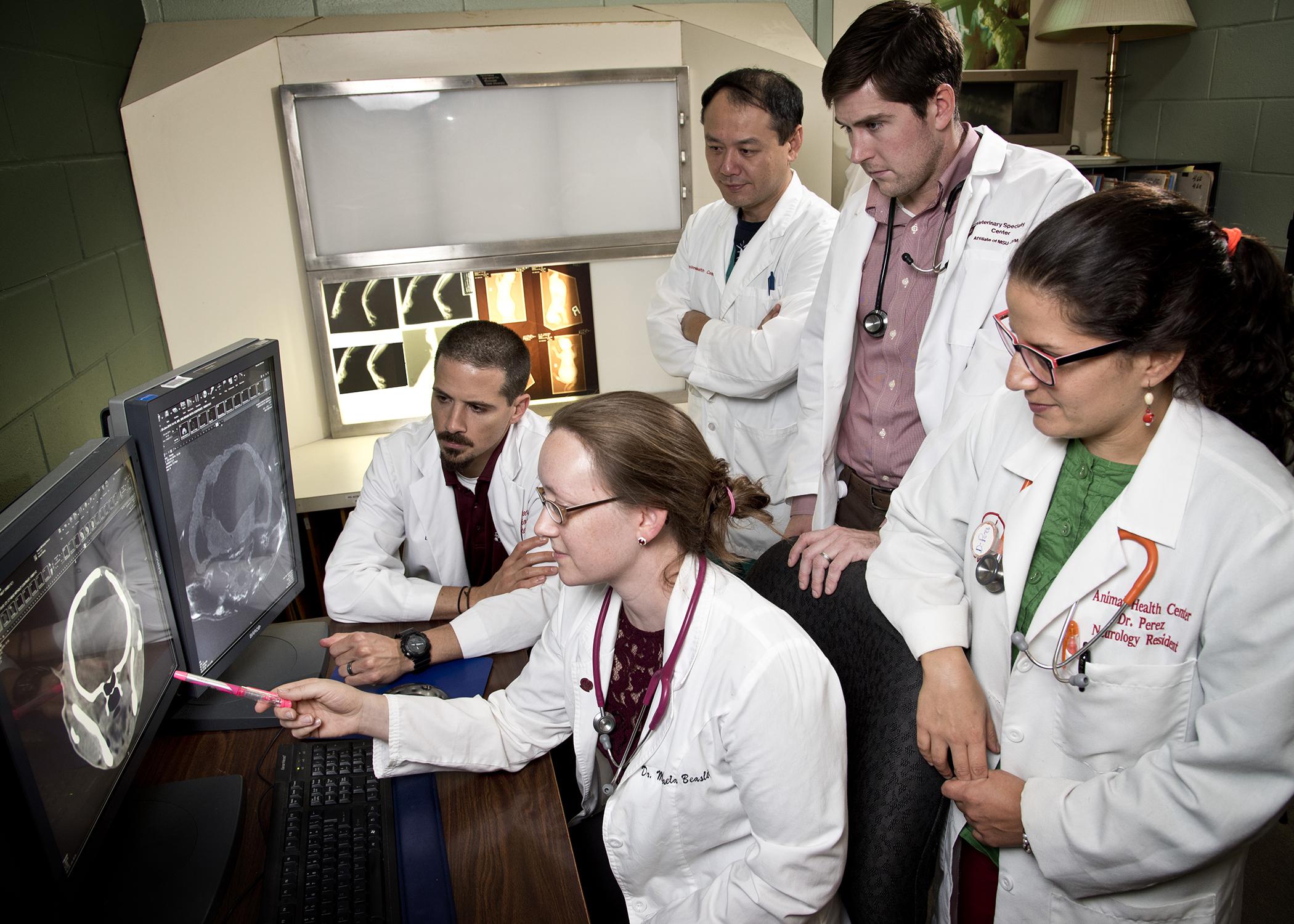
High-resolution imaging, including computerized tomography (CT) and magnetic resonance imaging (MRI) helps surgeons determine the best site to perform the procedure. Shores said the modified procedure has been successful in five patients, including a 10-week-old kitten and a 14-week-old puppy.
“Follow-up CT and MRI scans confirmed the new outlets were unobstructed and the ventricles had reduced in size,” he said. “Their clinical signs also had resolved or improved and remained improved over a period of six months.”
The MSU veterinary neurosurgeons who developed the procedure have presented the technique at two national conferences and hope that the veterinary medical community will adopt it as a viable method of managing hydrocephalus and reducing surgical complications, Beasley said.
Contact: Karen Templeton, 662-325-1100


