Embryo Transfer in the Beef Herd
Most female breeding cattle produce one calf per year. Embryo transfer (ET) allows a producer to quickly multiply the genetics of the top females in the herd. When artificially inseminated with semen from bulls with high genetic merit, these top females produce calves with superior genetics. Females in the herd with less desirable genetics can serve as recipients for the embryos, and the overall genetic quality of the herd may be drastically improved in a short amount of time.
The first calf from a transferred embryo was born in the early 1950s using a surgical procedure. Today, embryos can be collected and transferred without surgery, allowing this reproductive management tool to become more common, especially in the seedstock segment of the beef industry. As of 2008, approximately 1.6 percent of beef cattle operations in the United States used embryo transfer. While the results of superovulation and embryo transfer vary, producers can take steps to increase the probability of success.
Embryo transfer requires two components: 1) generating and obtaining (flushing) the embryos from the donor female, and 2) transferring each embryo into a different female (recipient), which gestates and gives birth to that fetus. These two components do not necessarily have to be done by the same producer. Embryos can be produced and sold to other producers who transfer them into their own recipient females.
Selecting a Donor Female
Selection of each donor female is one of the most important decisions in ET. Donor females should be of superior genetic worth and marketability to justify ET costs. Mating decisions should be made considering the genetic worth and economic value of potential calves. The reproductive potential of a donor female must also be acceptable.
The ideal donor female has had regular estrous cycles beginning at a young age, routinely conceives with no more than two inseminations, maintains a 365-day or less calving interval, calves without difficulty, is free of reproductive abnormalities and disease, and has no conformational or known genetic defects. Good nutritional management of these females is critical for productivity as embryo donors. This involves managing body condition and providing proper nutrients, including minerals, important to reproductive function.
Superovulation
Donor females are superovulated, which allows them to ovulate more than one oocyte. Combinations of hormones are administered over a matter of days. Embryo transfer technicians often have their own preferred protocol, but an example of a superovulation protocol is depicted in Table 1.
|
Day |
Time |
Amount/type of injection |
|---|---|---|
|
1 |
AM |
3.5 cc FSH |
|
PM |
3.2 cc FSH |
|
|
2 |
AM |
2.8 cc FSH |
|
PM |
2.4 cc FSH |
|
|
3 |
AM |
2.2 cc FSH |
|
PM |
1.9 cc FSH |
|
|
4 |
AM |
1.7 cc FSH and 5 cc Prostaglandin F2α |
|
PM |
1.5 cc FSH and 5 cc Prostaglandin F2α |
|
|
5 |
AM |
0.9 cc FSH |
|
PM |
0.7 cc FSH |
|
|
6 |
AM |
Donor should display estrus; artificially inseminate |
|
PM |
Artificially inseminate |
|
|
7 |
AM |
Artificially inseminate |
If a group is to be superovulated, they need to be synchronized so that they are at the same stage of the estrous cycle. An estrous synchronization protocol, such as the Select Synch CIDR® protocol, is frequently used to synchronize the estrous cycle (Figure 1). This involves giving an injection of gonadotropin-releasing hormone (GnRH) and inserting a CIDR® vaginal insert (Controlled Internal Drug Release device; Pfizer Animal Health, New York, NY). The GnRH synchronizes the next follicular wave, and the CIDR® releases progesterone, which prevents ovulation from occurring while it is inserted. Seven days later, the CIDR® is removed, and an injection of prostaglandin F2α (PGF2α) is given, which causes the corpus luteum (CL) on the ovary to regress and allows the cow to begin estrus. This estrus, called the “marker heat,” is indicative of her ability to ovulate and signals that she is properly synchronized. Only females showing this marker heat should continue in the protocol.
Nine to eleven days after the marker heat, a series of injections of decreasing amounts of follicle-stimulating hormone (FSH) are administered. This encourages multiple follicles to grow, which leads to the ovulation of multiple oocytes. Following the FSH injections, two injections of PGF2α are administered 12 hours apart. Estrus detection begins, and females are typically inseminated with one straw of semen at the initiation of estrus; two straws 12 hours later; and again with one straw 24 hours after the initiation of estrus (Figure 1). This provides an adequate number of live sperm to fertilize all oocytes shortly after ovulation. Embryos are flushed 7 days after estrus.

Embryo Collection
A series of steps are taken to collect embryos from a donor female. A technician first injects a numbing agent (usually Lidocaine) into the spinal cord, near the tailhead, to eliminate the muscular contractions that normally occur in the rectum. This allows the technician to more easily manipulate the uterus and flushing tubing in the reproductive tract. A catheter, which is placed over a metal stylette, is inserted through the cervix, similar to passing a rod used for AI (Figure 2). Once the tip of the catheter is in the uterine body, a bubble, or cuff, is inflated to help keep the catheter in place and prevent fluid from leaking out of the uterine body.


The metal stylette is then removed, and tubing is attached to the catheter to connect it to the bag of flushing media (Figure 3). Flushing media is allowed to flow into the uterine body, where the technician will massage the uterus to allow the fluid to “pick up” any embryos present. The fluid is then allowed to flow out of the uterus and through a very fine filter that traps the embryos.
The technician repeats this process several times, sometimes focusing on one uterine horn and then the other. Once complete, the cuff on the end of the catheter is deflated, and the tubing and catheter are removed. An injection of PGF2α is administered so that if any embryos are missed in the collection process, they will not develop a pregnancy in the donor cow.
Embryo Processing, Grading, and Freezing
The filter used to capture the embryos is rinsed to collect them in a dish. This dish is placed under a microscope so the technician can locate and retrieve each individual embryo. Once all embryos are found, each receives a two-number classification according to developmental stage and quality (grade).
The first number designates the developmental stage the embryo has achieved and ranges from one (unfertilized oocyte) to nine (expanding hatched blastocyst). The second number designates the quality and ranges from one to four, with one being the best quality. Typically, embryos with a quality of one or two can be either transferred fresh or frozen for later use. Those with a quality of three are much less viable after freezing so are only transferred fresh. Embryos with a quality score of four are generally not transferred at all.
Bovine Embryo Development Stages
Stage 1: unfertilized
Stage 2: 2- to 12-cell
Stage 3: early morula
Stage 4: morula
Stage 5: early blastocyst
Stage 6: blastocyst
Stage 7: expanded blastocyst
Stage 8: hatched blastocyst
Stage 9: expanding hatched blastocyst
Bovine Embryo Grades
Grade 1: excellent or good
Grade 2: fair
Grade 3: poor
Grade 4: dead or degenerating
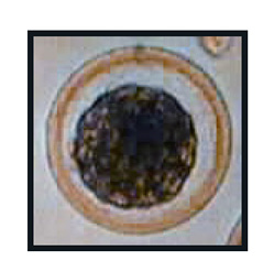
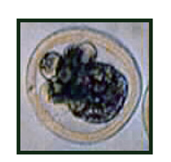
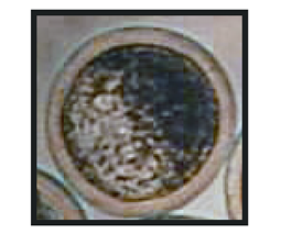
Figure 4 shows embryos with grades of 4-1, 4-3, and 6-1. The 4-1 embryo is very tightly compacted compared to the 4-3. The 4-3 has more cells and fragments outside the compacted morula, which is an indication of poorer quality. Embryo technicians are trained to detect slight differences in embryos and grade them accordingly.


Figure 4. From top to bottom, embryos with grades of 4-1, 4-3, and 6-1 (International Embryo Transfer Society Manual, 4th edition).
Embryos are now ready to be transferred or frozen for later use. Around eight transferable or six freezable embryos per flush can be expected, on average. However, some females will produce fewer embryos and others can produce 30 or more quality embryos in one flush.
If embryos are to be frozen, they are washed and stored in a cryoprotectant. This fluid minimizes the damage done to an embryo when it is frozen. Ethylene glycol is used most often because it enables the direct transfer of embryos from the straw in which they were frozen. Occasionally, embryos stored and frozen in glycerol are encountered. These must be thawed by a trained embryologist using a microscope and thawing media.
Embryos are loaded into 0.25 cc plastic straws and labeled. The label for each embryo must include the technician’s code (assigned by the International Embryo Transfer Society), cow breed, dam and sire registration numbers, the number of embryos (more than one embryo can be stored in each straw), and the date.
The straws are then placed into a special freezer used for embryos. These particular freezers decrease the temperature in a slow, controlled fashion, which is necessary for embryo viability. Once frozen, the straws are placed in labeled goblets and canes (like those used to store semen) and plunged into liquid nitrogen in a storage tank. Embryos can be stored indefinitely as long as the tank and liquid nitrogen are carefully maintained.
Transferring Embryos
When embryos are to be transferred fresh, they are loaded directly into 0.25 cc plastic straws and transferred using a rod similar to an AI rod. Most AI rods are designed to use 0.5 cc straws, but those used to inseminate with sex-sorted semen are designed for 0.25 cc straws. To thaw frozen embryos, the straw should be removed carefully from the tank, held in the air for 10 seconds, and then placed in a water bath between 77 and 86 °F (25–30 °C), which is cooler than the temperature used to thaw semen. The straw should remain in the water bath until any ice has melted, approximately 30 seconds. The cap is removed from the straw and loaded into the transfer rod, and then a transfer sheath is placed over it. The rod is passed through the cervix, and the embryo is deposited in the uterine horn on the same side that ovulation occurred, as determined by the presence of a CL on the ovary.
Recipient Females
In anticipation of having fresh embryos available for transfer, recipient females should be in good body condition and health, on a proper plane of nutrition, and on a sound herd health program. Synchronize them so that they are in the same place in their estrous cycle as the donor female. Use the Select Synch protocol (Figure 1) or any other protocol that is designed to allow for timed AI (refer to Mississippi State University Extension Service Publication 2614 Estrus Synchronization in Cattle).
Time the start of the synchronization protocol so that both the donor and the recipients will be at day 7 of the estrous cycle when flushing occurs. This will allow the fresh embryos to be transferred directly to a recipient that has a uterus ready for the establishment of pregnancy. If a recipient does not have a CL at the time of transfer, she should not be used. Therefore, more females should be set up as recipients than the number of expected embryos to ensure there are enough good recipient candidates available. Pregnancy rates average approximately 65 percent when fresh embryos are transferred into recipients at the correct stage of the estrous cycle.
Another option is to freeze the embryos and transfer them into recipients at a more convenient time. This can be done when an insufficient number of recipient females are available at the time of flushing, if more embryos were collected than was expected, if the ideal calving date is not 9 months after embryo flushing, or if embryos are being sold or transported to another location. A recipient female can receive an embryo 7 days after she is detected in standing estrus, or a group of recipients can be synchronized and multiple embryos transferred at one time. The freezing process slightly reduces the viability of embryos. Therefore, pregnancy rates are approximately 55 percent when transferring frozen-thawed embryos.
Embryo Transfer Services
Because successful ET programs require highly trained technicians, be diligent when selecting people to perform these services. Embryos may not be marketable unless they have proper documentation, such as freeze codes. Some breed associations require reporting of embryo removal dates or other information on calves resulting from embryo transfer to be eligible for breed registration. Technicians should complete certificates of embryo recovery, freezing, or transfer as appropriate. Many technicians are members of the International Embryo Transfer Society. These embryologists develop reputations for proficiency among producers, so it is useful to visit with other producers using ET services to locate a desirable technician.
The cost of ET services is highly variable. There may not be a qualified technician available in the local area or at the particular time needed, so considerable travel may be required for an on-farm visit from a technician. Travel expense is often included in the bill to the producer. Some ET facilities allow donor females and/or recipient females to be delivered to the facilities for embryo collection and transfer services. It is wise to schedule embryo transfer services several months in advance.
Conclusion
Superovulation and embryo transfer are used to increase the number of offspring from genetically outstanding females as well as superior sires. The superovulation procedure causes more oocytes to be ovulated than is normally the case, and these oocytes are fertilized using AI. Seven days after insemination, embryos are collected from the donor by flushing the uterus, and these embryos are transferred into recipient females to gestate the embryos until birth. For more information on cattle reproduction or related topics, contact an office of the Mississippi State University Extension Service.
References
U.S. Department of Agriculture. 2009. National Animal Health Monitoring System BEEF 2007-08. Washington, D.C.
The information given here is for educational purposes only. References to commercial products, trade names, or suppliers are made with the understanding that no endorsement is implied and that no discrimination against other products or suppliers is intended.
Publication 2681 (POD-07-21)
Revised by Brandi Karisch, PhD, Associate Extension/Research Professor, Animal and Dairy Science; from an earlier edition by Jane A. Parish, PhD, Professor and Head, North Mississippi Research and Extension Center.
The Mississippi State University Extension Service is working to ensure all web content is accessible to all users. If you need assistance accessing any of our content, please email the webteam or call 662-325-2262.


